Brodmann area 33
Image
See also
Navigation menubirnlex_176668630
Brodmann area 33
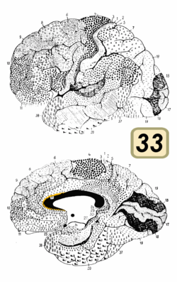 Brodmann area 33 (shown in orange)
 Medial surface of the brain with Brodmann's areas numbered.
Details
Identifiers
Latin
Area praegenualis
NeuroLex ID
birnlex_1766
FMA
68630
Anatomical terms of neuroanatomy[edit on Wikidata]
Brodmann area 33, also known as pregenual area 33, is a subdivision of the cytoarchitecturally defined cingulate region of cerebral cortex. It is a narrow band located in the anterior cingulate gyrus adjacent to the supracallosal gyrus in the depth of the callosal sulcus, near the genu of the corpus callosum. Cytoarchitecturally it is bounded by the ventral anterior cingulate area 24 and the supracallosal gyrus (Brodmann-1909).
Image
Animation.

Medial view.
See also

Wikimedia Commons has media related to Brodmann area 33.
- Brodmann area

This neuroanatomy article is a stub. You can help Wikipedia by expanding it.

Brodmann area 33 (shown in orange)

Medial surface of the brain with Brodmann's areas numbered.
NeuroLex ID
Anatomical terms of neuroanatomy
[edit on Wikidata]
Image

Animation.

Medial view.
See also
This neuroanatomy article is a stub. You can help Wikipedia by expanding it.


